Interdepartmental microscopy platform
All the information on the website: https://piattaformadimicroscopia.unimib.it/
The Interdepartmental Microscopy Platform aims to bring together under a single structure the interdepartmental services of optical and electronic microscopy. An electronic microscopy service has been active at the University of Milan Bicocca since 2004 and in 2007 it became part of the Microscopy Platform.
The Platform was born from the collaboration of the Departments of:
The headquarters of the Microscopy Platform is located in the reserved spaces on floor -2 of building U3 in Piazza della Scienza 2.
The Microscopy Platform offers the possibility to use the service to all the staff of the University of Milano-Bicocca and other Universities, to the staff of public research centres for their institutional activities, as well as to the staff of non-profit centres. Access to the microscope is also open to private individuals in the form of commissioned research. All services are regulated by regulations and a price list.
Instruments
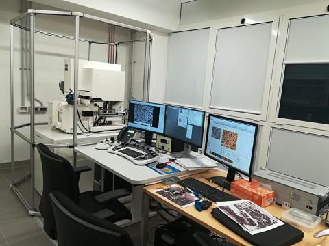
As far as electron microscopy is concerned, the instruments allocated are the following:
- FEG microscope (SEM-FEG) Gemini 500 Zeiss complete with QUANTAX EDS 4000, EBSD and STEM microanalysis system
- Microscope Focused Ion Beam (FIB) QUANTA200 3D FEI
- Variable pressure scanning electron microscope (SEM) Tescan VEGA TS 5136XM, equipped with EDAX microanalysis
- Transmission electron microscope (TEM), maximum acceleration voltage 110kV (instrument available from the Department of Environmental and Earth Sciences)
The electron microscopy equipped with EDX probe allows:
-the 'morphological' analysis of a sample at much higher magnifications than those allowed by optical microscopy, with a very high contrast. In this way and by means of various expedients, such as, for example, the possibility of using secondary electrons or 'backscattered' electrons, it is possible to analyse the sample in 3D, enhancing the surface or volume components of the portions observed.
-analyzing the chemical elements present in each point of the sample under observation, also making 'mappings' of chemical elements in more or less large areas.
The transmission microscope (TEM) allows even higher resolutions than SEM but requires proper sample preparation.
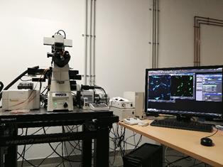
For optical microscopy in the microscopy platform is allocated the Nikon A1R Confocal Microscope.
Thanks to the Nikon A1R confocal optical confocal microscope, with the possibility of classical (galvanometric) and Resonant (for acquisitions with high temporal resolution) scanning, it is possible to perform the imaging of isolated biological structures or in situ, with considerable improvement in contrast and spatial resolution compared to classical optical microscopy techniques.
The microscope is also equipped with a camera for epifluorescence acquisition and a microincubator for experimental measurements with CO2 and temperature control.
Director
Prof. Giancarlo Capitani (giancarlo.capitani@unimib.it)
For information on:
SEM and TEM microscopy on materials and geological samples: Giancarlo Capitani (giancarlo.capitani@unimib.it)
SEM and TEM microscopy on biological samples: Paride Mantecca (paride.mantecca@unimib.it)
Optical Microscopy: Marcella Rocchetti (marcella.rocchetti@unimib.it)
To book the service:
Dr. Paolo Gentile (paolo.gentile@unimib.it ); Tel. +39 02 6448 2017
Dr. Tiziano Catelani (tiziano.catelani@unimib.it); Tel. +39 02 6448 2914